ALL ARTICLES AND PRODUCT INFORMATION PROVIDED ON THIS WEBSITE ARE FOR INFORMATIONAL AND EDUCATIONAL PURPOSES ONLY. The products offered on this website are furnished for in-vitro studies only. In-vitro studies (Latin: in glass) are performed outside of the body. These products are not medicines or drugs and have not been approved by the FDA to prevent, treat or cure any medical condition, ailment or disease. Bodily introduction of any kind into humans or animals is strictly forbidden by law.
How does Epitalon enhance sleep while protecting DNA and Telomeres?
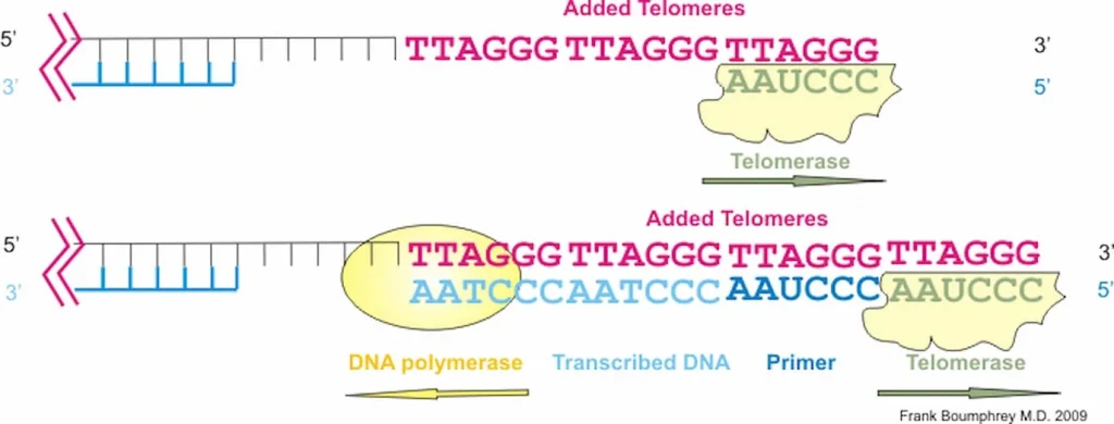
Epithalon Peptide Induces Telomerase Activity and Telomere Elongation in Human Somatic Cells and Overcomes the Hayflick Limit
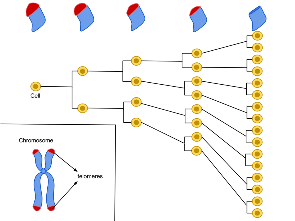
Each cell contains DNA as an instruction manual for how to divide and grow. The DNA inside of each cell is shielded by proteins called telomeres. During cellular division, a new cell must take some telomeres from its originating cell to shield the DNA of the new cell. The telomeres shorten after every cell division because the new cell can only take a portion of the telomeres from the previous cell, else the previous cell’s DNA will become completely unprotected.Once there are no left over telomeres to take, the cell stops dividing. This happens after a single cell divides and grows about 64 other cells, which is known as the Hayflick limit. This limit exists because cells without shield material are more vulnerable DNA damage. If the DNA of a cell becomes damaged, the cell will follow broken instructions. If the instructions within the DNA of the cell are damaged, then the cell may not be able to eliminate itself through the process of apoptosis like it is supposed to.”The telomere length is increased by approximately 33% in epitalon treated cells [by increasing the telomerase enzyme that strengthens telomeres].” (10)“Telomerase is a reverse transcriptase that has two distinct functions, to replicate pre-existing chromosome ends (telomeres) and to heal broken chromosomes by de novo addition of telomeric sequences directly on to non-telomeric DNA.” (11)
“Addition of Epithalon to aging cells in culture induced elongation of telomeres to the size comparable to their length during early passages. Peptide-treated cells with elongated telomeres made 10 extra divisions (44 passages) in comparison with the control and continued dividing. Hence, Epithalon prolonged the vital cycle of normal human cells due to overcoming the Hayflick limit.” (12)
Peptides for Anti-Aging Research
Best Peptides for Anti-Aging Research
Anti-aging research has hit its stride in the last decade with advancements coming at an ever-faster pace. Recently, at Kyoto University in Japan, biologist Shinya Yamanaka has shared the Nobel Prize in medicine for work he did into how peptides can be used to reprogram adult stem cells to reduce the effects of aging. The research revealed that a protein cocktail is able to turn back the clock in mouse models, restoring the epigenomes of the mice to a more youthful state and reducing inflammation, musculoskeletal dysfunction, cognitive decline, and more[1].The advances made by Dr. Yamanaka come as no surprise to experienced anti-aging researchers because the trend has been toward more complex interventions using multiple peptides and approaches. According to Harvard’s David Sinclair, a founding member of the Paul F. Glenn Center for the Biology of Aging. According to Dr. Sinclair, the field has advanced during his career from extending the lives of relatively simple organisms like yeast and worms to being able to reverse aging in complex organisms like mice and non-human primates. Much of that progress has come from combining various techniques and layering knowledge to gain ever more sophisticated control of cellular aging. While Dr. Sinclair’s goal has primarily been to slow the onset of certain diseases associated with age, he points out that there is little difference between slowing disease onset and slowing aging. In fact, he points out that slowing the onset of heart disease, something that is commonly achieved in medicine today, is just an early form of slowing the aging process. Stem cell research and anti-aging proteins are just the next step on the path toward slowing the onset of aging and, eventually, maybe even halting aging altogether.
Peptides and Aging
At the forefront of current anti-aging research are techniques designed to reverse epigenetic changes in DNA. Epigenetic changes refer to changes in DNA expression patterns and not to changes in the DNA sequence itself. Research shows that, as part of the aging process, some genes are turned on and some are turned off. This leads to several the effects we associate with aging such as lower testosterone, lower growth hormone, changes in estrogen levels, alterations in wound healing, a decline immune function, changes in skin structure, alterations in learning and memory, and more.
The reversal of epigenetic changes has been shown in previous research to diminish or even reverse the effects of aging but has been difficult to achieve outside of cell culture. The discovery that peptides, particularly small peptides, can penetrate cell membranes and act as epigenetic signals has led to a rush to understand how these peptides can be useful both in isolation and in combination. Dr. Yamanaka’s research focused on the ability of certain peptides, in combination, to slow or reverse certain aspects of aging. There are a handful of peptides that have proved to be particularly interesting because of their widespread and obvious effects on aging in a variety of tissues. These peptides have helped to not only reverse the effects of aging in mouse models but have revealed some of the mechanisms by which aging occurs. Thus, these peptides, discussed below, have helped to advance the science a great deal. What is more, they are still offering insight to scientist who design careful trials to explore their function and mechanisms of action.
How Do Telomeres Impact Aging and Autophagy?
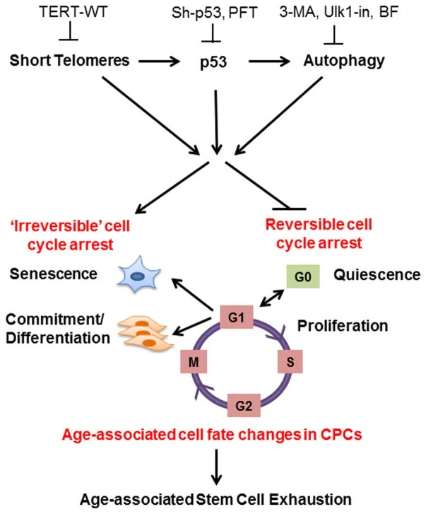
Loss of Telomere Leads to p53 and Autophagy Induced Cell Death.
“autophagy-deficient cells … continued to proliferate and bypassed crisis” (2)
“In summary, cells in telomere crisis undergo cell death through autophagy, which is triggered by chromosome breakage and transduced by the cGAS–STING pathway. As cell death in crisis represents the final barrier against neoplastic transformation, a cancer therapy that involves inhibition of autophagy could be counterproductive… Moreover, cells lacking either cGAS or STING proliferated beyond crisis. “
“Autophagy mediates the turnover of cytoplasmic macromolecules to support cellular homeostasis. Autophagy generally blocks apoptosis, but in specific circumstances it can lead to cell death through excessive degradation of cell constituents. The authors studied telomere crisis using human fibroblasts and epithelial cells, in which the RB and/or p53 pathways were suppressed; these cells bypassed senescence and entered replicative stress, exhibiting telomere attrition, chromosome fusions and cell death.”
“Telomere deprotection through TRF2 depletion was sufficient to activate autophagy independently of replicative crisis, and genetic suppression of telomere fusions in TRF2-depleted cells reduced the accumulation of cytosolic DNA and attenuated autophagy, suggesting that fusion-dependent cytosolic DNA is required for the telomeric autophagy response. ” (2)
“The cell fate of CPCs changes with age and is characterized by a switch away from proliferation and quiescence (reversible form of cell cycle arrest) towards senescence and increased basal commitment (‘irreversible’ forms of cell cycle arrest) accounting for age-associated stem cell exhaustion. Mechanistically, short telomeres activate p53 that induces autophagy and at least partially contributes to the age-associated change in cell fate. Blunting telomere shortening via overexpression of TERT-WT, silencing p53 , or treating with pharmacological inhibitors of p53 (PFT) and autophagy (3-MA, Ulk1-In, BF) selectively attenuate senescence and basal commitment and reverse cell fate of aged CPCs.” (3)
Circadian Rhythm Controls Telomeres and Telomerase Activity. “Circadian clocks are fundamental machinery in organisms ranging from archaea to humans. Disruption of the circadian system is associated with premature aging in mice, but the molecular basis underlying this phenomenon is still unclear. In this study, we found that telomerase activity exhibits endogenous circadian rhythmicity in humans and mice. Human and mouse TERT mRNA expression oscillates with circadian rhythms and are under the control of CLOCK–BMAL1 heterodimers.
CLOCK deficiency in mice causes loss of rhythmic telomerase activities, TERT mRNA oscillation, and shortened telomere length. Physicians with regular work schedules have circadian oscillation of telomerase activity while emergency physicians working in shifts lose the circadian rhythms of telomerase activity. These findings identify the circadian rhythm as a mechanism underlying telomere and telomerase activity control that serve as interconnections between circadian systems and aging.” (4)
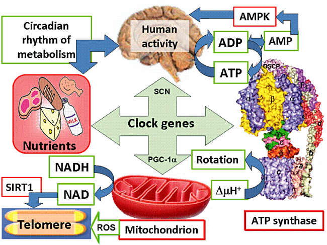
“Human activity is driven by NADH and ATP produced from nutrients, and the resulting NAD and AMP play a predominant role in energy regulation. Caloric restriction increases both AMP and NAD and is known to extend the healthspan (healthy lifespan) of animals. Silent information regulator T1 (SIRT1), the NAD-dependent deacetylase, attenuates telomere shortening, while peroxisome proliferator-activated receptor γ coactivator 1α (PGC-1α), a master modulator of gene expression, is phosphorylated by AMP kinase and deacetylated by SIRT1. Thus, PGC-1α is a key component of the circadian oscillator that integrates the mammalian clock and energy metabolism.
Reactive oxygen species produced in clock mutants result in telomere shortening. The circadian rhythms produced by clock genes and lifestyle factors are ultimately controlled by the human brain and drive homeostatic and hedonic feeding and daily activity. ” (9)
Telomerase and TERT mRNA expressions exhibit endogenous circadian rhythm.
Human and mouse TERT mRNA expression are under the control of CLOCK–BMAL1 heterodimers.
CLOCK deficient mice have shortened telomere length and abnormal oscillations of telomerase activity and TERT mRNA.
Emergency physicians working in shifts lose the circadian rhythms of telomerase activity.
What is Epithalon?
Epithalon is currently being studied and researched by Scientists and Doctors specializing in the field of anti-aging care and medicine. EPITHALON (Epitalon) is one of the most important breakthroughs in the study of anti-aging.
The Epithalon (Epitalon) tetrapeptide has been discovered by researchers in Russia. This was seen to reactivate the production of cell telomerase thus slowing down the aging process and rejuvenating the entire body. The development of molecular biology required bio-chemical studies that were nothing short of profound. Scientific work by Gobind Khorana and Marshall Nirenberg for many years resulted in defining codons or nucleotides and triplets and the genetic code of each of the 20 amino acids. This resulted in a Nobel Prize award in 1968 with Robert Holley. Nucleic acid investigations and identification of DNA and RNA base sequences were also conducted by the 1980 Nobel Prize winner for Chemistry Frederick Sanger along with Walter Gilbert and Paul Berg. These studies revealed the cause of aging.
Epithalon and Skin Rejuvenation
Epithalon and Skin Rejuvenation
Skin rejuvenation is often associated with wrinkles and lines, but the truth runs deeper than wrinkles. Skin becomes more fragile and thus more prone to damage as it ages. Damage to the skin compromises its protective barrier function and can increase risk of infection. Research into ways to strengthen skin can not only make skin look younger, but can protect people from serious medical conditions. Thus far, most skin rejuvenation research has focused on collagen and other large skin proteins. New research, however, suggests that short peptide molecules, like epithalon, may hold more promise in preserving and even rejuvenating skin.
Epithalon Overview
Epithalon (a.k.a. epitalon), is a short (just four amino acids long) peptide that has been demonstrated to have anti-aging and anti-cancer properties in rodent studies. Because epithalon is so short, it can penetrate the cell membrane, without the aid of transporters, and make its way to the nucleus of cells. This is important because, once in the nucleus, epithalon can affect the regulation of genes, activating some and deactivating others to cause cell-wide changes1.
Previous research has indicated that epithalon can stimulate immune system function that has been lost due to natural aging. Investigation of the mechanism of this action uncovered the ability of the Ala-Glu-Asp-Gly peptide chain (Epithalon) to interact with the promoter region of the interferon gamma gene. By promoting the production of interferon gamma, a key immune regulator, epithalon is able to boost functioning in T-cells and thus overall immunity and well being1,2.
The idea that short peptides might be able to affect DNA-level processes has caused a boom in the investigation and research of epithalon and other short peptides in animal models. Those investigations have led to the understanding that epithalon can impact skin aging by activating cellular repair processes, which often go dormant as we age.